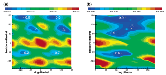
Redox-active tyrosine residues play important roles in long-distance electron reactions in enzymes such as prostaglandin H synthase, ribonucleotide reductase, and photosystem II (PSII). Spectroscopic characterization of tyrosyl radicals in these systems provides a powerful experimental probe into the role of the enzyme in mediation of long-range electron transfer processes. Interpretation of such data, however, relies critically on first establishing a spectroscopic fingerprint of isotopically labeled tyrosinate and tyrosyl radicals in nonenzymatic environments. In this report, FT-IR results obtained from tyrosinate, tyrosyl radical (produced by ultraviolet photolysis of polycrystalline tyrosinate), and their isotopologues at 77 K are presented. Assignment of peaks and isotope shifts is aided by density-functional B3LYP/6-311++G(3df,2p)//B3LYP/6-31++G(d,p) calculations of tyrosine and tyrosyl radical in several different charge and protonation states. In addition, characterization of the potential energy surfaces of tyrosinate and tyrosyl radical as a function of the backbone and ring torsion angles provides detailed insight into the sensitivity of the vibrational frequencies to conformational changes. These results provide a detailed spectroscopic interpretation, which will elucidate the structures of redox-active tyrosine residues in complex protein environments. Specific application of these data is made to enzymatic systems.